Malocclusions: Understanding and Managing Malocclusions in Dogs and Cats
Original document by Dr. Fraser A. Hale, DVM, FAVD, Dipl. AVDC. Edited and updated by Dr. Regan L. Morris, DVM, Dipl. AVDC
Malocclusions refer to misalignments of teeth and jaws. While pets don’t require perfect alignment, a functional and pain-free bite is essential for their health. This document outlines the anatomy of primary and adult teeth, classifications of malocclusions, reasons for treatment, and strategies for intervention and management.
Primary Dentition and Malocclusion Basics
Anatomy of Primary Teeth
- Dogs: The primary dental formula is 2x (i3/3, c1/1, p3/3), meaning each quadrant has three incisors, one canine, and three premolars. Notably, there are no primary molars or first premolars.
- Cats: The primary formula is 2x (i3/3, c1/1, p3/2). Cats lack primary molars, first premolars, and mandibular second premolars.
- Confusingly, the 3rd primary premolar in each quadrant has the structural anatomy and the function of the adult 4th premolar and the 4th primary premolars have the form and function of the adult 1st molars.
In both species, the developing adult teeth erupt in predictable locations relative to the primary teeth. For example, adult incisors erupt lingual or palatal to the primary incisors.
Classification of Malocclusions
Unless stated otherwise, the next session refers to primary teeth.
Normal Occlusion
Normal Occlusion: proper craniofacial development with appropriate jaw-length relationships (upper and lower jaws proper lengths in their own right and in relation to each other), proper jaw width relationship and all teeth present and in their proper position/alignment.
We see six upper and six lower incisors. The maxillary midline is directly above the mandibular midline and there is good symmetry right-to-left. The lower incisors are just behind the upper (scissors occlusion). The width of the mandibles is also appropriate compared to the maxilla.
The lower canine tooth is centred in the space between the upper third incisor and canine teeth and is tipped labially at enough of an angle that it is not in direct contact with the maxillary soft tissue
Class 1: Dental Malocclusion
- Description: Normal craniofacial structure, but one or more teeth are misaligned.
- Example: Linguoversion (aka “base-narrow”) of the canines, where lower canines are displaced lingually and cause trauma to the palate. This is painful – think about how painful it is when puppies play bite too hard with their needle sharp teeth! It can damage the underlying adult teeth and even be severe enough to cause an oronasal fistula!
- Treatment:
- Immediate extraction of the offending primary tooth to alleviate pain and create a path for the proper eruption of the adult tooth.
- Ball therapy at this age WILL NOT HELP. However, after extraction of the deciduous tooth, owners need to initiate “ball therapy”as soon as the lower adult canine teeth start to emerge through the gingiva at 5.5 to 6 months of age. Ball therapy refers to the use a properly sized ball to encourage the eruption of adult canines at the correct angle. Think of it as a removable orthodontic appliance.
Class 2: Skeletal Malocclusion
- Description: The mandible is too short compared to the maxilla, creating an “overbite” (Note that “overbite” is not correct terminology). Note the wide gap between the upper and lower incisors and how the lower canines are now trapped to the palatal side of the upper canine teeth. This is a serious skeletal malformation!
- Issues:
- Lower canines may impinge on the palate, causing pain and trauma.
- Abnormal dental interlock may restrict mandibular growth.
- Treatment:
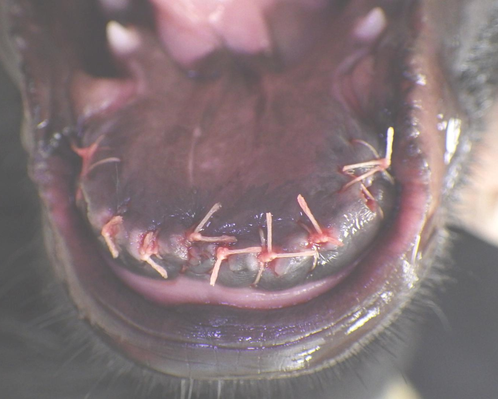
- Extraction of lower primary incisors and canines to eliminate trauma and mechanical restrictions on mandibular growth.
- Note: Genetic limitations may prevent full correction, and additional treatment for adult teeth may be necessary.
Craniofacial development is under genetic control and virtually all malocclusions are genetic and heritable. One possible exception would be a “wry-bite” or facial asymmetry secondary to a known trauma during development. In the absence of a known trauma that could reasonably be expected to have caused the deformity, all malocclusions should be considered genetic and heritable. Affected individuals should not be bred and neither should their parents. It might even be best to avoid breeding their litter mates.
Class 3: Reverse Skeletal Malocclusion
- Description: The maxilla is shorter than the mandible (“underbite”). This is common in brachycephalic breeds (e.g., Bulldogs). Sadly, this serious craniofacial abnormality is considered breed standard for a great many breeds of dog and several breeds of cats (the brachycephalics) despite this anatomy being associated with a great many problems.
- Issues:
- Crowded and rotated upper teeth predispose to periodontal disease.
- Traumatic contacts between lower incisors and soft tissue.
- Under-eruption of certain teeth, leading to gum inflammation and infection.
- Treatment:
- If the patient is not of brachycephalic breeding and the malocclusion is mild, then removal of some primary teeth to alleviate any abnormal interlocks might be worthwhile.
- If the patient is a member of a brachycephalic breed (or is a mix with a brachycephalic parent) and the teeth are not causing trauma, there may be no point in doing anything with the primary teeth.
- Remove primary teeth if they are causing trauma to the soft tissues (these are painful to the patient).
- Owners of brachycephalic breeds should anticipate significant dental care needs for adult teeth, typically requiring professional intervention.
Class 4: Wry Bite
- Description: Asymmetry between the right and left sides of the jaw, often caused by uneven growth.
- Treatment:
- Extract teeth causing trauma or impeding growth.
- Severe cases may require specialized orthodontic or surgical interventions.
Dental Interlock: Explanation and Importance
Dental interlock refers to the natural interdigitation of teeth between the maxilla (upper jaw) and mandibles (lower jaw), which helps maintain proper jaw alignment during growth. In normal occlusion, this interlock ensures that as one jaw grows, the other follows, maintaining the correct jaw length relationship. For example, during a maxillary growth spurt, the upper canine teeth push against the lower canines, encouraging simultaneous mandibular growth. Similarly, during mandibular growth, the lower incisors push against the upper incisors and palatal structures, promoting coordinated maxillary development. This mechanism plays a critical role in achieving a balanced craniofacial structure.
However, abnormal dental interlock occurs when teeth are misaligned, impeding natural growth. For instance, in Class 2 malocclusions (short mandible), the lower canines may penetrate the palatal mucosa, creating an “abnormal interlock” that staples the mandible to the maxilla in this dysfunctional relationship. Even the lower incisors can be a problem as they can get hung up behind the palatal rugae and incisive papilla. This abnormal dental interlock can act as a mechanical impediment to the desirable growth of the mandibles. This mechanical restriction prevents the mandibles from catching up, potentially leading to further deformities. If the mandible attempts to grow but cannot move due to the interlock, it may bend ventrally, typically between the adult fourth premolar and first molar, exacerbating the issue. Similarly, in Class 3 malocclusions (short maxilla), the upper incisors can become trapped behind the lower incisors, creating another form of abnormal interlock that prevents the maxilla from catching up to the mandible.
Addressing abnormal interlocks early, such as through the extraction of problematic primary teeth, can alleviate mechanical restrictions and promote unimpeded growth. This intervention helps the animal express its genetic growth potential while minimizing pain and structural abnormalities. Dental interlock highlights the interconnected nature of craniofacial growth and underscores the importance of timely veterinary dental care.
The growth of the maxilla and the mandibles are under the control of different genes. The maxilla tends to grow faster initially but the mandibles soon catch up. Maintenance of a proper jaw-length relationship depends on a proper dental interlock.
Why Treat Malocclusions?
Malocclusions can cause significant pain, even if pets don’t show outward signs. Key reasons for treatment include:
- Relieving Pain: Misaligned teeth often traumatize soft tissues or other teeth, causing discomfort.
- Promoting Normal Growth: Correcting primary malocclusions helps ensure proper craniofacial development, maximizing the animal’s genetic potential.
- Preventing Future Complications: Early intervention minimizes the need for invasive and costly treatments later in life.
Treatment Options and Timing
Timing is Critical
- Early detection (at the first vet visit, 8–12 weeks of age) allows for timely extractions and interventions.
- Delays reduce the effectiveness of treatment and can result in permanent skeletal abnormalities.
- The longer surgery is delayed, the longer the pet has been in pain and the less time is had before the adult teeth erupt. The value of intervening diminishes with every week that goes by and drops off dramatically after 16 weeks of age. By 20 weeks, we have missed the boat and must deal with the problems the adult teeth create at 6 to 7 months of age.
Interventions for Primary Malocclusions
Extractions: Careful removal of primary teeth causing trauma or misalignment, under anesthesia, is the most common intervention. Extracting the primary teeth causing abnormal interlocks to promote normal growth, acknowledging genetic limits may persist.
Ball Therapy: Encourages proper eruption of ADULT CANINE TEETH in Class 1 (and select Class 2) malocclusions.
It is important to know that when the lower canines are palatal to the upper canines because the mandibles are too short, Ball Therapy will be of no use. Ball Therapy only works when the lower canines just have to tip out towards the lip and there is nothing blocking their path to a desirable location.
I have also found that Ball Therapy will not move already erupted teeth. It does however often work very well to guide the teeth AS THEY ARE ERUPTING into a more labial position.
Persistent Primary Teeth
- Persistent primary teeth should be removed immediately when adult teeth begin erupting. Two teeth occupying the same space can misalign adult teeth and cause trauma.
- Early removal allows adult teeth to erupt into proper alignment and prevents complications.
Adult Teeth Eruption: Understanding Placement
The eruption of adult teeth follows a predictable pattern relative to their primary precursors, which is crucial for assessing and managing dental development in puppies and kittens. These rules ensure veterinarians can anticipate the trajectory of adult teeth during the early stages of a pet’s life:
- Incisors: Adult incisors always erupt on the lingual (tongue-side) or palatal (roof-of-mouth side) relative to the primary incisors.
- Lower Canines: The adult lower canines erupt on the lingual side of the primary lower canines.
- Upper Canines: Adult upper canines erupt mesially (toward the midline) and slightly forward of the primary canines.
This predictable pattern allows veterinarians to identify abnormalities early. For instance, if a primary tooth persists, it can misdirect the eruption path of the adult tooth, leading to misalignment or crowding. Persistent primary teeth must be extracted promptly to prevent these issues and allow adult teeth to erupt into their correct positions.
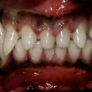
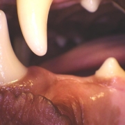
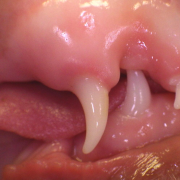
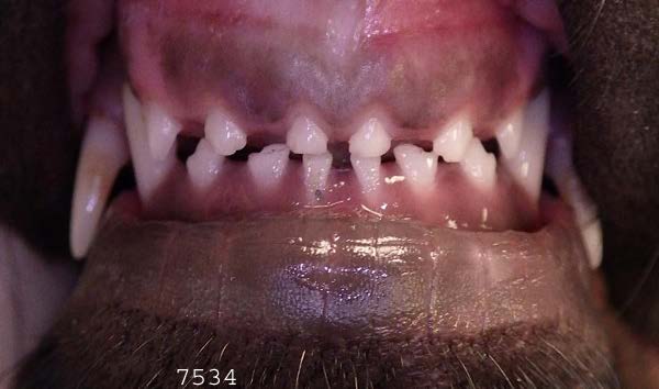
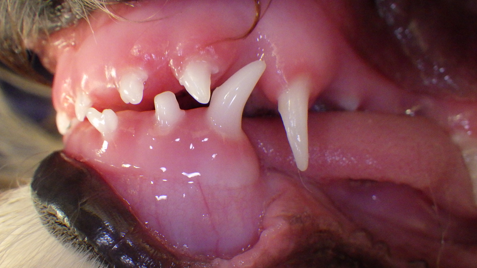
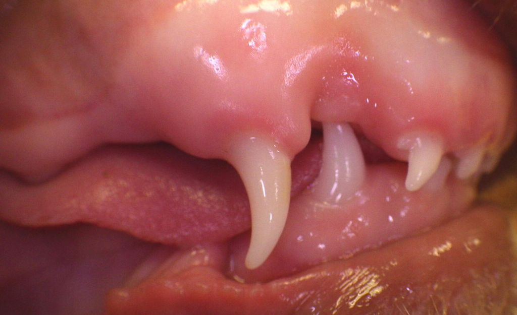
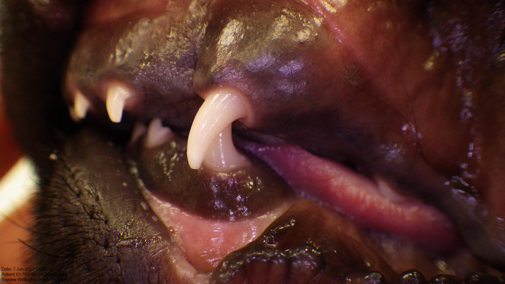
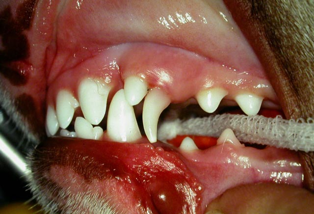
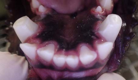
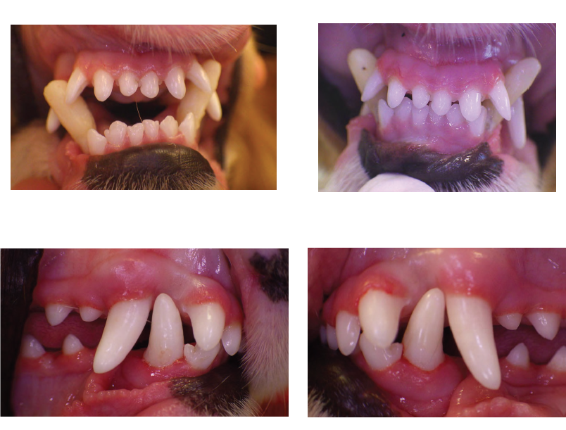
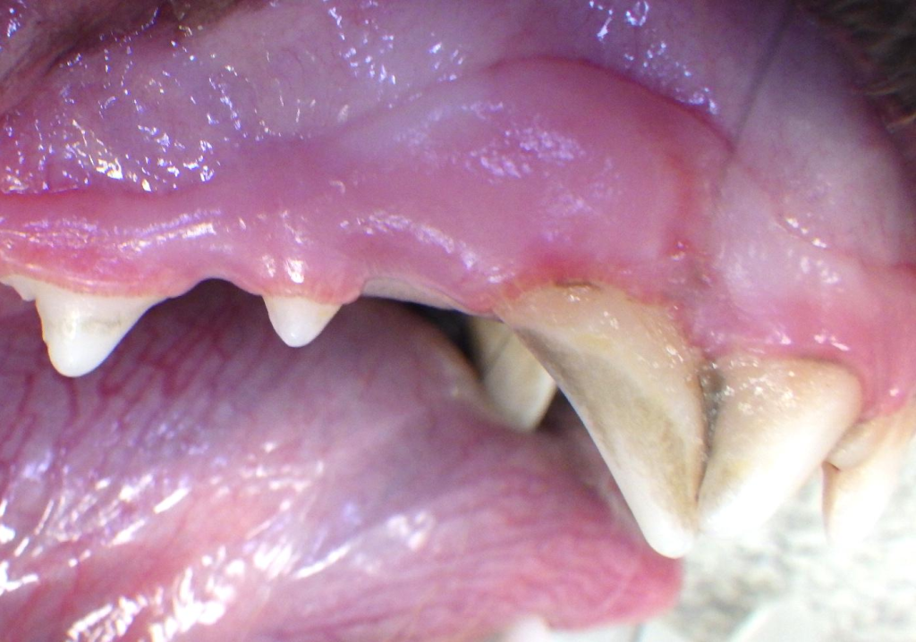
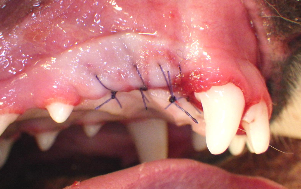
Note that the following images are not normal and are undesirable! All of these persistent primary teeth should be removed ASAP.
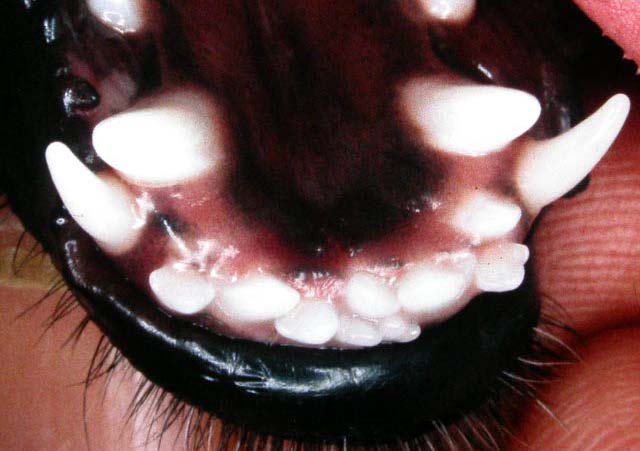
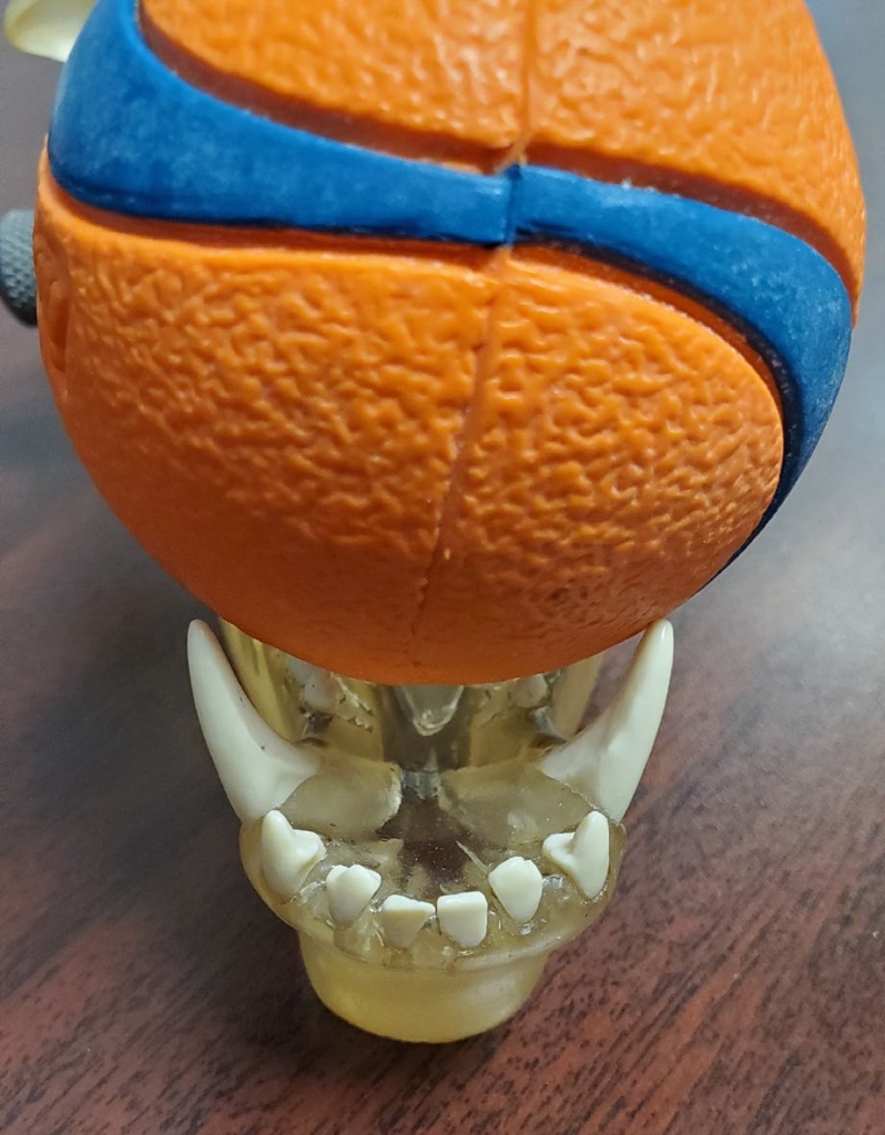
Early detection and intervention are vital to ensure that adult teeth align properly, avoiding complications like abnormal contacts, crowding, and trauma. Understanding where and how adult teeth erupt provides a foundation for managing both primary and mixed dentition phases in growing pets. In the third photo of this grouping, you will see that the upper left primary canine tooth is occupying the space meant for the erupting adult tooth. This is forcing the adult tooth to erupt too far forward in the mouth so there is too little space between it and the 3rd incisor (the space that is supposed to accommodate the crown of the lower adult canine). This will trap the lower adult canine tooth on the palatal side of the upper canine and force the lower canine into traumatic contact with the maxillary tissues. If the deciduous tooth is extracted immediately, the adult tooth will swing back into its proper location and allow for the lower canine tooth to swing labially/outward).
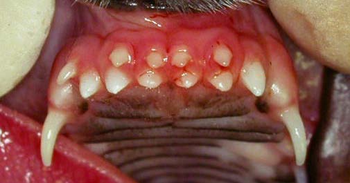
Similarly, if the lower primary canine tooth persists as the adult tooth is erupting (even if the primary had been properly positioned all along) the adult tooth, which will always be on the tongue side of the primary (second photo in this grouping) will be forced to erupt base narrow, driving it up into the palate. So, get the primary tooth out right away and if the jaw length relationship is normal or close to normal, adding Ball Therapy would also be a very good idea.
Management of Adult Malocclusions
Crown Reduction and Vital Pulp Therapy: Shortens or reshapes adult teeth causing trauma, often with pulp capping. This typically needs to be done at 7 months of age.
Orthodontic Appliance: Placement of Temporary Crown extensions to guide adult teeth into proper alignment. These require expertise and regular follow-up under anesthesia.
Extractions: In cases where malocclusions cause persistent trauma or crowding and is the most practical solution.
Special Considerations for Brachycephalic Breeds
Brachycephalic dogs often require extensive dental care due to congenital malocclusions and crowding. Treatment focuses on alleviating trauma, preventing periodontal disease, and improving oral function. Regular dental checkups and proactive care are essential.
Referrals and Advanced Care
Veterinarians should refer complex cases to specialists trained in veterinary dentistry and orthodontics. Steps include:
- Providing high-quality photographs and radiographs.
- Sharing detailed clinical history and test results.
- Encouraging owners to schedule appointments promptly to avoid missed opportunities for treatment.