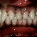
When you look at these photos, do you have any concerns for the pet?
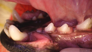
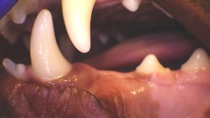
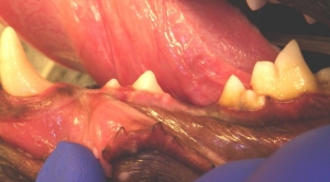
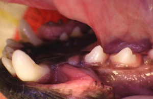
In the first two photos, we cannot see the first lower left premolar (tooth 305) and in the third photo, we cannot see the third lower left premolar (tooth 307).
How many teeth do dogs and cats have?
Dogs are supposed to have 42 permanent (adult) teeth and cats are supposed to have 30 permanent (adult) teeth. So why is it important that we make sure each and every one is accounted for? What are the consequences if the tooth is present, and we cannot see it?
There are several possibilities (the differential diagnoses) for a tooth that we cannot visualize when we look in a dog or cat’s mouth:
A missing tooth may be truly missing – ie. it never formed. This is referred to as hypodontia. Or it could have fallen out due to disease. But either way, if it is not present and the area is healthy, then it is no longer a concern.
A missing tooth could be a tooth that has fractured, and the roots remain under the gum. These roots would have been exposed to bacteria in the mouth (even if the gum has healed over top of them) and have a very good chance of having persistent infection (causing inflammation and pain to the pet). These roots need to be removed to resolve this. One exception may be if the tooth fractured due to replacement tooth resorption and the gingiva may be healthy and healed completely over the mostly resorbed tooth root(s). In this situation, it may be appropriate to leave it alone. That being said – if you are uncertain whether it is replacement resorption (vs other types of tooth resorption) then you cannot make this call. Leaving a resorbing tooth is also NOT appropriate if it is in a cat with Feline Chronic Gingivostomatitis.
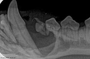
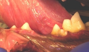
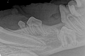
This first case is a four-year old 8 kg dog with an apparently missing first lower premolar (305). It is impacted and a cyst has destroyed the bone and compromised the blood supply and the supportive periodontal structures of the adjacent teeth. This dog has such a thin rim of cortex remaining in the area of the left mandible that it could have easily fractured with the smallest amount of force/trauma (ie. sneezing hard and banging its chin on the floor).
Post operative photograph and radiograph
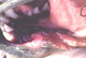

This next case is a one-year old 30 kg dog with much more robust bone however the cyst still destroyed the bone in the area and certainly left untreated, it could lead to jaw fracture. Thankfully the lower left canine tooth’s blood supply was still intact. If it had not been, then root canal or extraction would have been necessary. The vitality of this tooth was rechecked a few months later and was found to be sound. 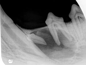
Any missing tooth in the mouth can be impacted, although some are more common than others. This last example is of a one-year old dog with an impacted third lower premolar.
The take home message here is that it is very important to account for every tooth in every mouth! Every pet that I meet, as part of the exam – I count their teeth and make sure they are all accounted for. If any are missing, and we don’t have knowledge for what happened to it, then we must investigate! These pets need a general anesthetic for intraoral imaging (radiographs) to determine if there is a problem or a potential problem.
For more reading on Dentigerous Cysts:
An overview of dentigerous cysts in dogs and cats – PMC (nih.gov)